Animal Responses
Divisions of the nervous system
The nervous system is divided into the central and peripheral nervous system. The central nervous system (CNS) consists of the brain and spinal cord whereas the peripheral nervous system (PNS) is made up of the nerves that connect the CNS to the rest of the body.
The PNS is then subdivided into the somatic nervous system, which controls conscious processes (e.g. running), and the autonomic nervous system which controls unconscious activities, such as breathing. The autonomic nervous system can also be subdivided into the sympathetic and parasympathetic branches. The sympathetic nervous system initiates the ‘fight or flight’ response which gears our body for action when sympathetic neurones release the neurotransmitter noradrenaline. The parasympathetic nervous system triggers the ‘rest and digest’ response. In this case, parasympathetic neurones release acetylcholine to calm the body down.

Brain structure
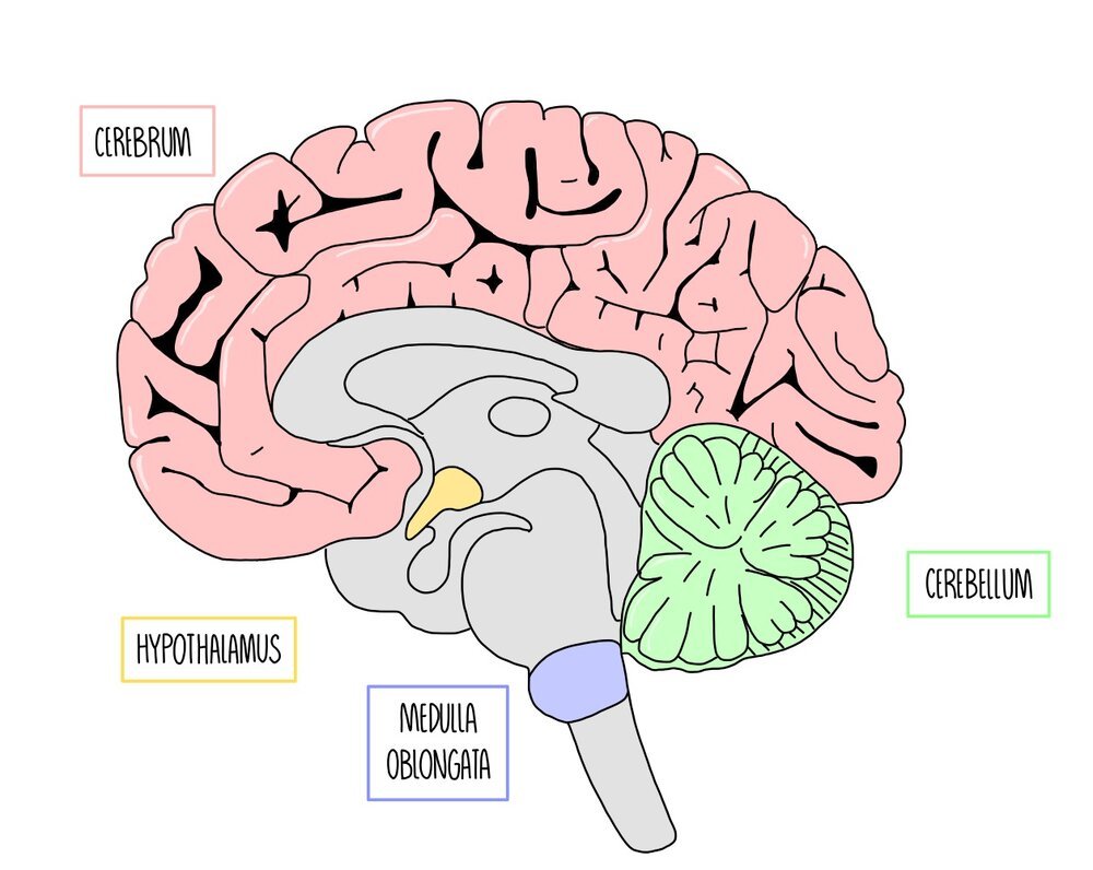
The brain can be divided into specific regions which have specific functions. Most of our brain is made up of the cerebrum, which is found at the top of the brain. It is divided into two cerebral hemispheres joined together by a band of nerve fibres called the corpus callosum. The thin, outer layer of the cerebrum is called the cerebral cortex. It is highly folded, giving it a really large surface area. The cerebrum is involved in ‘higher-brain functions’, such as processing language, vision, thinking and emotions.
Below the cerebrum is a structure called the hypothalamus, which is involved in homeostatic responses such as maintaining body temperature (thermoregulation). It also produces hormones that control the pituitary gland, which is found just beneath the hypothalamus.
The cerebellum is a leaf-shaped structure found towards the back of the brain. It is positioned underneath the cerebrum and is highly folded. It plays an important role in movement and balance. Things like learning to ride a bike or the movement involved in writing will involve a large input from the cerebellum.
Right at the base of the brain and above the spinal cord is a structure called the medulla oblongata. This is involved in unconscious processes, such as the regulation of breathing rate and heart rate.
Reflexes
Reflexes are rapid, unconscious actions that protect the body from harm. They bypass the conscious parts of our brain, which stops us from having to think about the action, which would waste time. Blinking and the knee-jerk response are two types of reflex action.
To prevent damage to the eye, you automatically blink if something touches it. Sensory receptors in the cornea are stimulated when something touches the eye, sending an electrical impulse along a sensory neuron to the central nervous system (CNS). In the CNS, a relay neuron passes the impulse between the sensory neuron and a motor neuron, which passes the impulse to the orbicularis oculi muscles in the eyelids. These contract and cause the eyelid to close.
If your quadriceps muscle stretches suddenly, you automatically straighten your leg to help keep your balance. Stretch receptors in the quadriceps muscle detect that the muscle is being stretched and send an electrical impulse along a sensory neuron to the spinal cord. In this case, the impulse is passed directly onto a motor neuron, which carries the impulse to the quadriceps muscle. The muscle contracts and causes the leg to straighten.
The fight or flight response
The fight or flight response prepares our body for action in response to a threat. It involves both hormones and nervous signalling.
Hormones – the pituitary gland releases a hormone called ACTH which acts on the adrenal glands, stimulating the release of steroid hormones (e.g. cortisol) from the adrenal cortex.
Nervous system – the sympathetic branch of the autonomic nervous system is activated which signals to the adrenal glands to release adrenaline from the adrenal medulla.
The release of steroid hormones and adrenaline from the adrenal glands has the following effects on the body:
Stimulates the breakdown of glycogen into glucose (glycogenolysis)
Redirects blood flow away from the digestive system and towards muscles and the brain (through vasoconstriction and vasodilation)
Increases heart and breathing rate
Causes erector pili muscles in the skin to contract, causing hairs to stand on end – for animals, this makes them look bigger (and scarier).
Control of heart rate
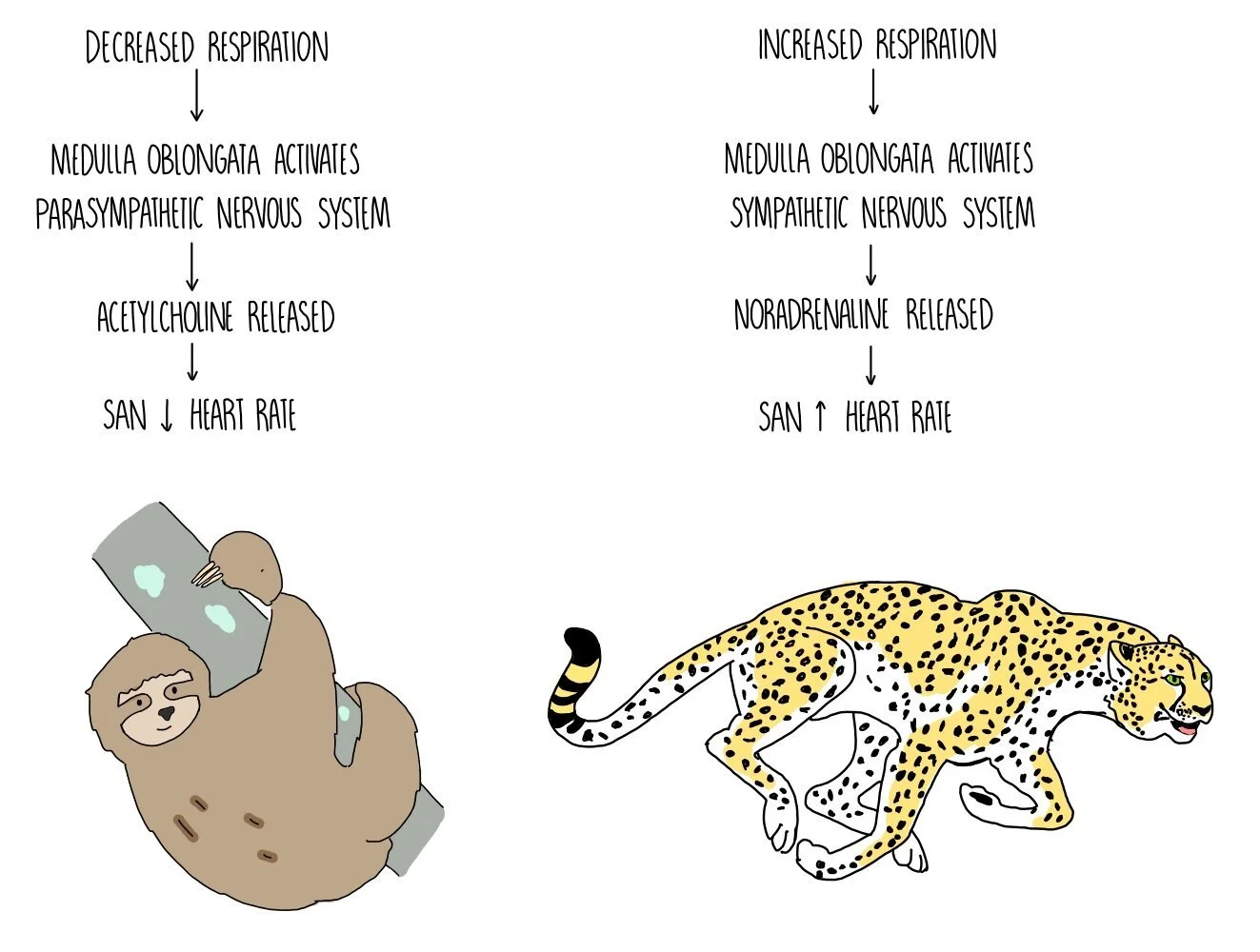
The medulla oblongata is a brain region found at the bottom of the brain, in the brain stem. It is involved in unconscious processes, such as controlling heart rate and breathing rate. A part of the medulla oblongata called the cardiovascular control centre is responsible for changing heart rate according to our body’s needs. It works by sending impulses along sympathetic or parasympathetic neurones which release different neurotransmitters onto the sino-atrial node (SAN) - the SAN then modifies its rate of firing to slow down or speed up the heart rate.
Two types of receptors, baroreceptors (pressure receptors) and chemoreceptors (chemical receptors) are responsible for detecting stimuli in the blood and signalling to the medulla oblongata to modify our heart rate. Baroreceptors detect changes in blood pressure and are found in the aortic and carotid bodies. Chemoreceptors detect the concentration of oxygen in the blood. They are also sensitive to changes in pH resulting from the carbon dioxide dissolved in the blood (its reacts with the water to form carbonic acid), which is an indication of oxygen availability. Chemoreceptors are also located in the aortic and carotid bodies.
When the medulla oblongata receives signals from baroreceptors and chemoreceptors, it changes the rate at which the SAN fires. If blood pressure or oxygen concentration is low, the cardiovascular control centre increases the rate of SAN firing through activation of the sympathetic nervous system. The sympathetic nervous system is involved in the ‘fight and flight’ response and increases heart rate through the release of a neurotransmitter called noradrenaline, which binds to receptors on the SAN. On the other hand, high blood pressure and high oxygen concentration causes the cardiovascular control centre to reduce the rate of SAN firing by activating the parasympathetic nervous system. This is involved in the ‘rest and digest’ response and decreases heart rate through the release of another neurotransmitter called acetylcholine. Acetylcholine binds to receptors on the SAN to slow down its rate of firing.
Student’s t-test
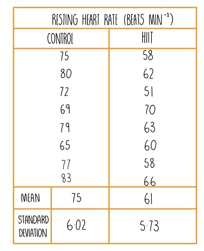
The Student’s t-test is used to determine whether the means for two sets of data are significantly different. For example, let’s say that you’re investigating whether an exercise program known as high intensity interval training (HIIT) is effective at lowering resting heart rate. Here’s our data for the two groups: the exercise group and the control group (no training).
To see if the treatment has had an effect on resting heart rate (i.e. that any differences are not just due to chance), we will need to carry out a student’s t-test. We do this by carrying out the following steps.
State the null hypothesis - this is always that there is no significant difference between the means of the two groups i.e. there is no significant difference between the resting heart rates of the group that completed the exercise program and those that didn’t.
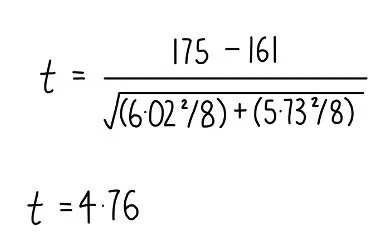
Use the formula below to calculate a value for t.
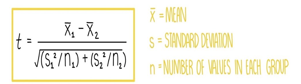
Putting in the mean values for each group, the standard deviations and the group size, we can work out a value for t as follows:
Compare your t value to the critical value. To work out the critical value from a table of critical values, you first need to work out how many degrees of freedom you have. The degrees of freedom is calculated by doing (n1 + n2) - 2. In our experiment, there is eight individuals in each group so we do (8 + 8) - 2 = 14. We then need to find the critical value at 14 degrees of freedom and a probability of 0.05 (5%). This means that were is a less than 5% chance that any differences between the two groups is due to chance.
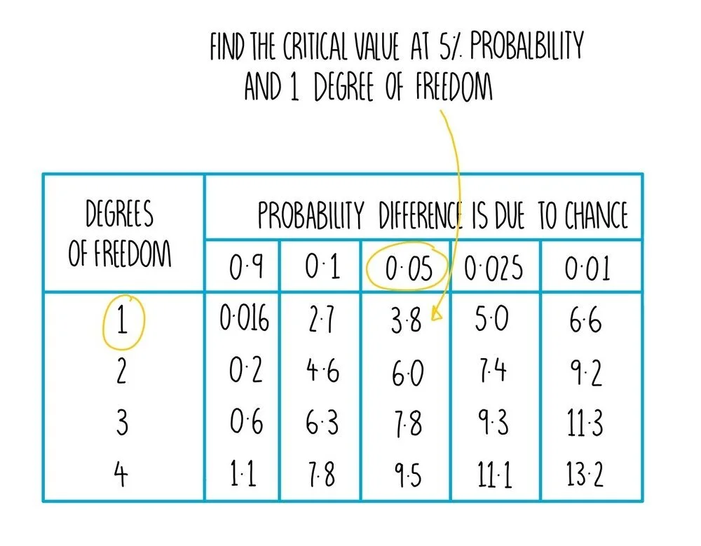
Determine whether the two means are significantly different - if the value obtained from the t-test is greater than the critical value at a probability of 5% or less, then you can be 95% confident that the difference between the two means is significant and not simply due to chance. You can reject the null hypothesis. In our example, our t-test value of 4.76 is greater than the critical value (2.14) so we can conclude that there is a significant difference in resting heart rate between individuals who underwent HIIT compared to the control group.
Structure of skeletal muscle
There are different types of muscle - for example, skeletal muscle, smooth muscle and cardiac muscle. Skeletal muscle is the type of muscle used for physical movement such as when we pick up objects or go for a run.
Skeletal muscle is made up of bundles of long muscle cells, called muscle fibres. The organelles inside muscle cells tend to have the prefix sarco- stuck to the front of their name. The cell membrane of muscle cells is called the sarcolemma and the cytoplasm of muscle cells is called the sarcoplasm. The sarcolemma folds into the sarcoplasm, creating something called transverse (T) tubules which help to spread electrical impulses throughout the cell. Muscle cells have a special organelle called the sarcoplasmic reticulum, which stores calcium ions for muscle contraction. Muscle cells also differ from other cells in that they contain many nuclei (they are multinucleate) and lots of mitochondria to generate ATP for muscle contraction. In addition, muscle fibres contain long cylinders of protein called myofibrils, which enable the muscle fibre to contract.
Sarcomeres
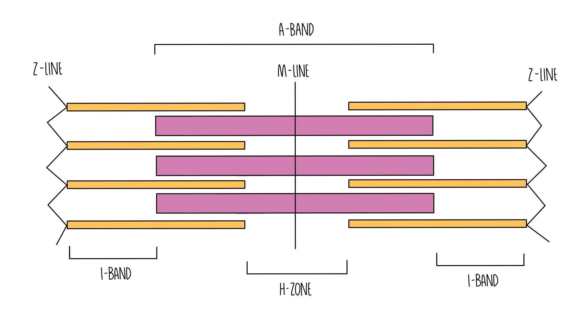
Myofibrils are made up of many short units called sarcomeres, which are made up of two types of myofilament: myosin and actin.
Myosin is a thick myofilament and appears as a dark band (called the A band) under the microscope.
Actin is a thin myofilament and appears as a light band (called the I band) under the microscope.
At the end of each sarcomere is a Z-line. Sarcomeres are joined together lengthways at the Z-line.
Right in the middle of the sarcomere is a region called the M-line.
The H-zone refers to the portion of the A-band which only contains myosin filaments (and not the portions where actin overlaps with myosin).
Sliding Filament Theory
When muscle fibres contract, the myosin and actin myofilaments move closer together by sliding over one another. This makes the sarcomere shorter and they contract. Remember that the actin and myosin myofilaments themselves don’t contract - they always stay the same length. As the muscle fibre relaxes, the myofilaments slide away from each other and move further apart, lengthening the sarcomere.
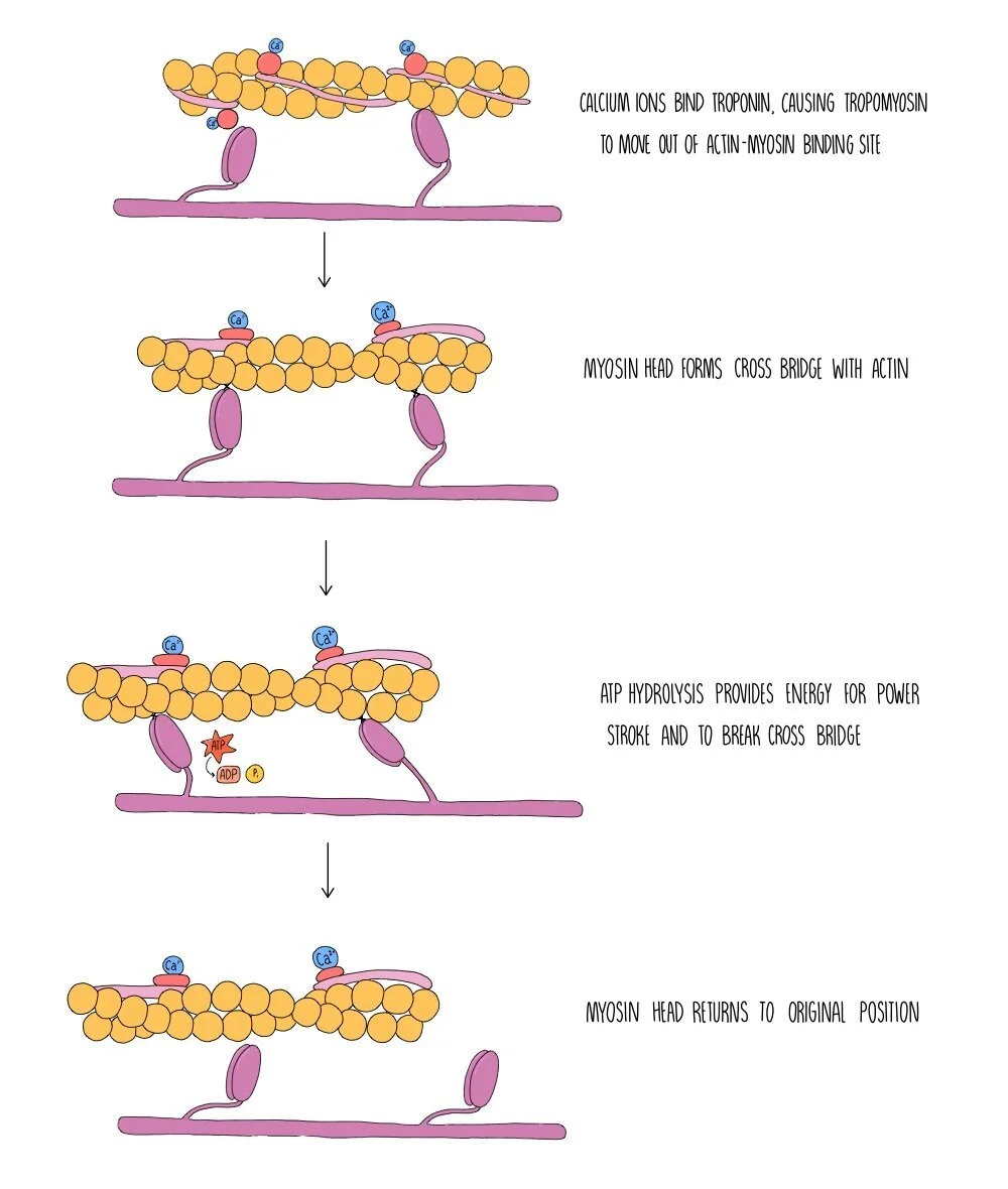
Myosin myofilaments possess head groups which contain binding sites for actin and ATP. Myosin heads are a globular and hinged, allowing them to move back and forth and enabling it to slide the actin filaments closer towards it. The region on actin where myosin binds is called the actin-myosin binding site. This binding site is blocked under resting conditions by two proteins called tropomyosin and troponin. When the muscle is not contracting, tropomysin covers the actin-myosin binding site and is held in place by troponin.
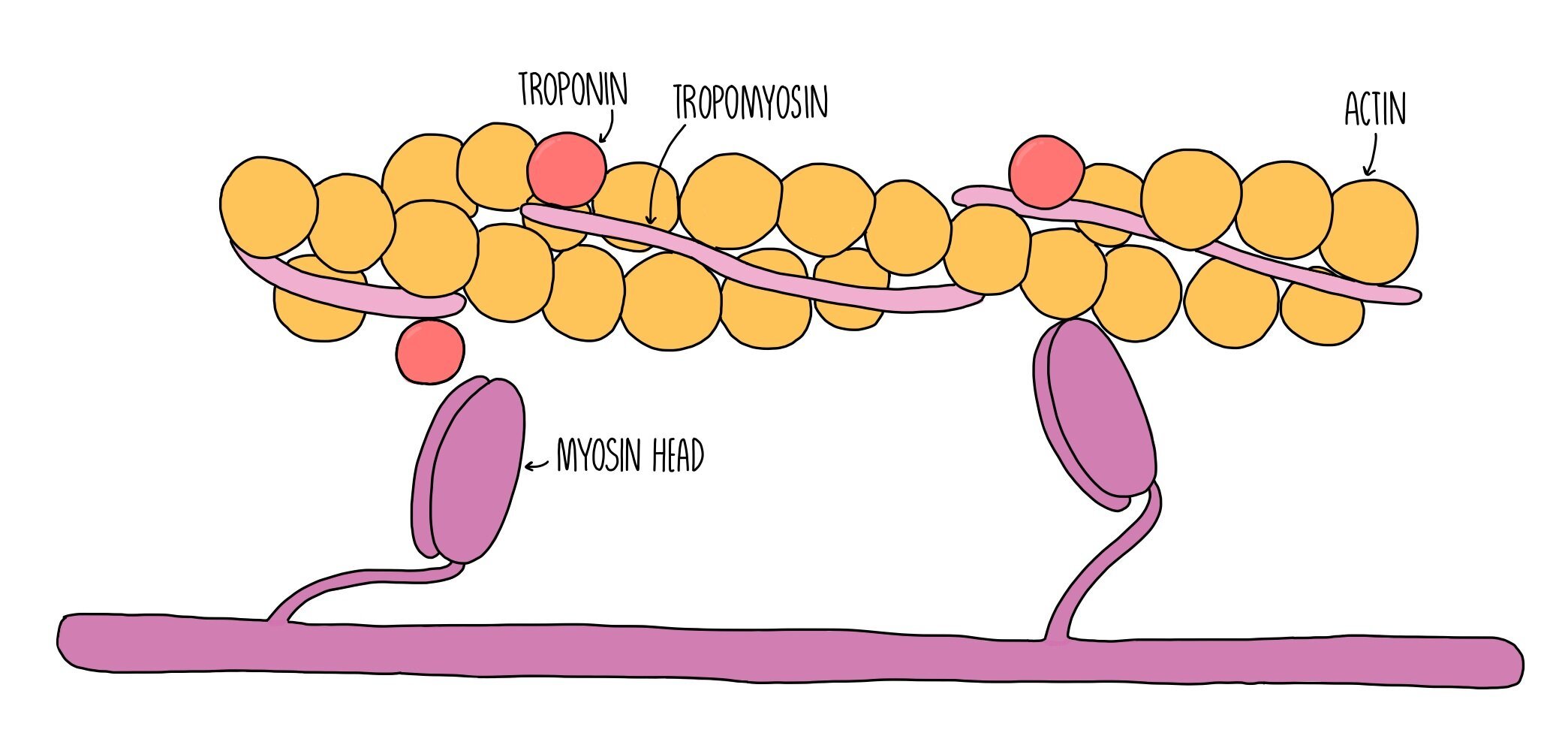
When an action potential (nerve impulse) arrives at a muscle fibre, a wave of depolarisation passes along the sarcolemma and down the T-tubules. This stimulates the sarcoplasmic reticulum to release calcium ions, which bind to troponin. Binding of calcium ions to troponin cause it to change shape, which pulls the tropomyosin out of the actin-myosin binding site. Now that the binding site is uncovered, the myosin head can bind to actin forming a bond called an actin-myosin cross bridge.
The release of calcium ions also activates the enzyme ATPase which catalyses the hydrolysis of ATP into ADP and inorganic phosphate. The energy released from ATP hydrolysis is used by the myosin head group to move backwards, pulling the actin filament closer towards it in a sort of rowing action which is referred to as a power-stroke. ATP hydrolysis also provides the energy to break the actin-myosin cross bridge. The myosin head can then reattach to a binding site further along the actin filament. The process is repeated, pulling the actin further and further towards the myosin filament. This shortens the sarcomere and results in muscle contraction.
When the muscle stops being stimulated, calcium ions move back into the sarcoplasmic reticulum by active transport, in a process which also requires ATP. The troponin molecules reform their original shape, pushing tropomyosin back into the actin-myosin binding site. Myosin can no longer bind to actin and the actin myofilaments slide back to their original position. The sarcomere lengthens and the muscle becomes relaxed.
Sources of ATP
Muscle contraction requires ATP for the power stroke, breaking the cross-bridge and for actively transporting calcium ions back into the sarcoplasmic reticulum once muscles have finished contracting. The ATP can be generated in three different ways:
Aerobic respiration
Oxidative phosphorylation generates most of the ATP in aerobic respiration.
This takes place in the mitochondria.
Can only take place when oxygen is plentiful e.g. low-intensity exercise.
Anaerobic respiration
Small amounts of ATP are produced in glycolysis.
Takes place in the cytoplasm.
Produces lactic acid which leads to muscle fatigue.
Suitable for short bouts of high-intensity exercise.
Phosphocreatine
In muscles, ATP be made by taking a phosphate group from phosphocreatine and phosphorylating ADP.
In this reaction, phosphocreatine is converted into creatine, which is removed from the body via the kidneys.
Phosphocreatine is stored in muscle cells and can generate ATP very quickly (but soon runs out).
Used for very short bursts of vigorous exercise.
It is anaerobic (does not use oxygen) and alactic (does not produce lactic acid).
Neuromuscular junctions
Neuromuscular junctions (NMJs) are the fancy name for a synapse between a motor neuron and a muscle cell. NMJs use the neurotransmitter acetylcholine and contain receptors called nicotinic cholinergic receptors. Unlike typical synapses:
The postsynaptic membrane in NMJs is folded into clefts which store the enzyme acetylcholinesterase, which breaks down acetylcholine.
Acetylcholine always acts as an excitatory neurotransmitter.
The postsynaptic membrane contains a higher number of receptors.
Types of muscle
There are three types of muscle: skeletal muscle (aka voluntary muscle), smooth muscle (aka involuntary muscle) and cardiac (heart) muscle. They have the following properties:
Skeletal muscle:
Striated (striped) appearance
Under conscious (voluntary) control
Contains fast-twitch fibres which contract quickly but fatigue quickly and slow-twitch fibres which contract and fatigue more slowly – fast-twitch are used for rapid movements (like sprinting) while slow-twitch are used for posture and endurance sports
Muscle fibres have multiple nuclei and can be several centimetres long
Smooth muscle with different tissue layers highlighted. Credit: Laird Sheldahl (Wikipedia)
Smooth muscle:
Cells are spindle-shaped (pointy at the ends) with no stripes
Under unconscious (involuntary) control
Contract slowly and do not fatigue
Each fibre has a single nucleus and is around 0.2 mm long
Found in blood vessels and in the walls of internal organs
Cardiac muscle:
Contains cross-striations (striped appearance) – but not as many as skeletal muscle
Fibres are connected by intercalated discs – these have low resistance so electrical activity easily passes between fibres
Fibres are branched at the ends
Contracts on its own accord (myogenic) but the rate of contraction is controlled by the autonomic nervous system.
Contracts rhythmically and does not fatigue.
Each fibre has a single nucleus and is around 0.1 mm long
Found in the walls of the heart
Measuring muscle fatigue using electromyography
You can measure the muscle fatigue by recording the electrical activity of skeletal muscles. The electrical activity represents the brain telling the muscles to move – the greater the electrical signal, the more tired the muscle is, since the brain is having to recruit more muscle fibres to help lift the weight.
Method:
Attach two electrodes to the muscle e.g. biceps
Attach a third electrode on a part of the body that isn’t moving – this is the control
Turn off any electrical equipment in the room to reduce ‘noise’ in the signal
Connect the electrodes to an amplifier – this increases the electrical signal to make it easier to read. Connect the electrodes and amplifier to a computer.
A straight line should appear on the electromyogram when the muscle is relaxed.
Fluctuating lines will appear when the muscle contracts e.g. flexing the biceps
Lift a weight – the amplitude of the lines increases with time because more motor units are required to lift the weight. As the muscle fatigues, the amplitude increases further as even more motor units are recruited to lift the weight.