Cell Division
Mitosis and the Cell Cycle
Mitosis is a type of cell division where cells produce identical copies of themselves and is used for growth and repair and asexual reproduction. It differs from meiosis, which is the type of cell division used to produce gametes.

Mitosis occurs as part of the cell cycle which consists of four distinct phases. First, interphase takes place which is made up of three growth phases (called G1 phase, S phase and G2 phase), followed by mitosis.
Gap Phase 1 (G1) - cell grows bigger and replicates its organelles. A high amount of protein synthesis is taking place in order to build new organelles.
Synthesis Phase (S) - the cell replicates its DNA
Gap Phase 2 (G2) - the cell keeps growing until all of the organelles have duplicated.
Once the DNA has replicated, each chromosome now consists of two sister chromatids, connected by a structure called the centromere. The mitochondria produce more ATP which will provide the energy for cell division and the ribosomes will be synthesising a high level of proteins to replicate organelles.
There are two ‘checkpoints’ in the cell cycle - one before S phase and one straight after S phase. During these checkpoints, the cell is checking its DNA for errors. This minimises the chances of duplicating any mutated DNA into the replicated cell.
The Stages of Mitosis
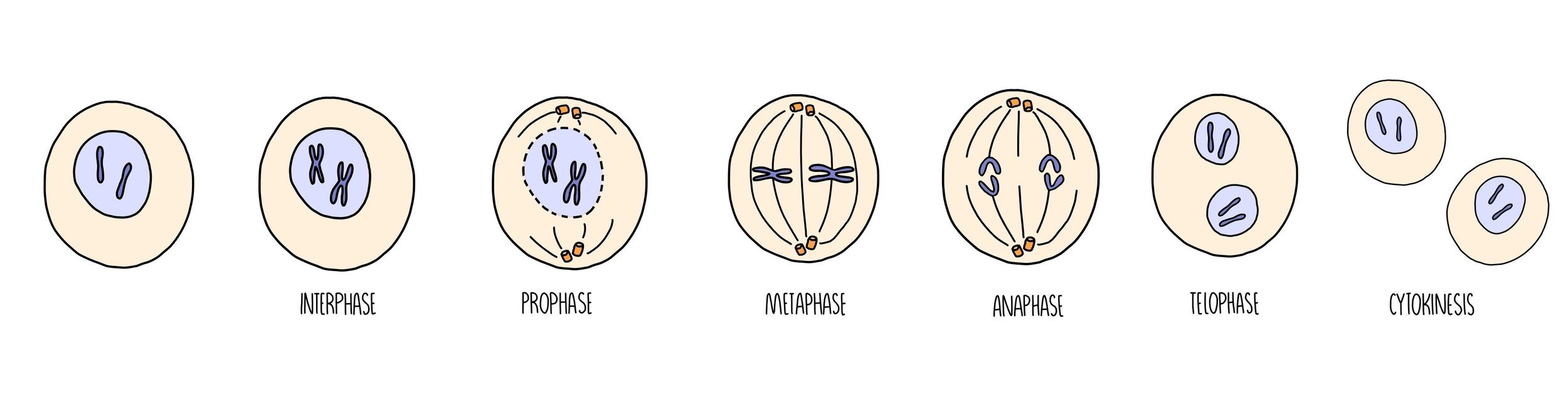
Mitosis can be divided into a series of stages depending on what’s going on with the chromosomes in the cell. You can use the acronym PMAT (pass me another tequila) to help you remember the order.
Prophase - the chromosomes condense (they become shorter and fatter) and the nuclear envelope disintegrates. The centrioles move to opposite poles of the cell and form spindle fibres.
Metaphase - the chromosomes line up along the middle of the cell. They attach to the spindle fibre by their centromere.
Anaphase - the centromere splits and the chromatids are pulled to opposite poles of the cell.
Telophase & cytokinesis - the two groups of chromsomes decondense (they become long and thin) and a nuclear envelope reforms around them, forming two new nuclei. The cytoplasm divides (cytokinesis) and the plasma membrane pinches off to form two new, genetically-identical cells.
Investigating mitosis in squashed root tips
You can see mitosis happening in root tip cells by staining the chromosomes and observing under the microscope. We use cells right from the tips of the roots because this is where mitosis is taking place (in the meristem tissue).
Method:
Cut a thin section of tissue from the tip of a growing root.
Pipette a set volume of 1M hydrochloric acid into a boiling tube and place in a 60oC water bath.
Place the plant tissue in the boiling tube and leave for five minutes.
Rinse the root tip with cold water and dry using a paper towel.
Cut the root tip so that you have a thin layer of cells (about 2 mm) and spread out onto a microscope slide using a mounted needle.
Add a drop of Toluidine blue O stain to the tissue and place a cover slip on top. Push down on the cover slip to squash the cells and allow light to pass through. Be careful not to push sideways otherwise the chromosomes will become damaged.
Use a light microscope to visualise the cells and identify the stages of mitosis. Any cells with visible chromosomes will be undergoing mitosis (as the chromosomes are condensed).
Gametes
Gametes are sex cells (the sperm and egg in humans). Gametes are haploid which means they contain half the number of chromosomes as the rest of the cells which make up our body. This means that when two gametes fuse during sexual reproduction, the fertilised egg (called a zygote) contains the full number of chromosomes i.e. it is diploid. In humans, the diploid number of chromosomes is 46 (23 pairs), which means that gametes contain just 23 chromosomes.
During sexual reproduction, the nucleus of the sperm cell fuses with the nucleus of the egg cell - this fusion of nuclei is called fertilisation.
Meiosis
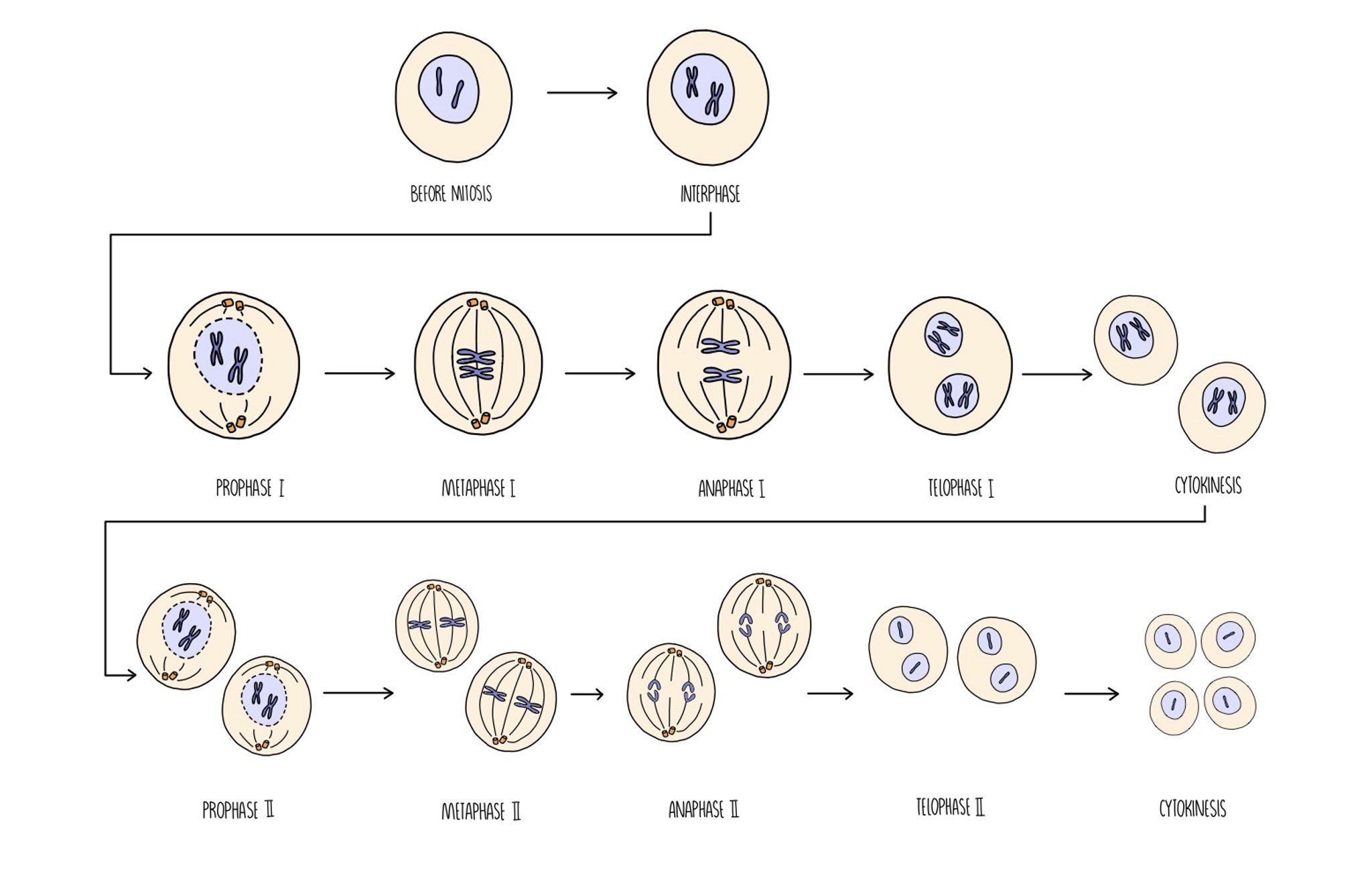
Meiosis is the type of cell division which produces gametes for sexual reproduction. Unlike mitosis, the daughter cells are genetically different from the parent cell and contain just half the number of chromosomes (i.e. they are haploid). When two haploid gametes join during fertilisation, a diploid cell called a zygote is formed. Meiosis involves two rounds of cell division which are referred to as meiosis I and meiosis II.
It takes place in the following stages:
Meiosis I
Interphase: the DNA replicates so there are now two identical copies of each chromosome (referred to as chromatids).
Prophase I: chromatids condense and arrange themselves into homologous pairs (called bivalents). Crossing over occurs (see below). The nuclear envelope disintegrates and spindle fibres form.
Metaphase I: homologous chromosomes line up along the equator and attach to the spindle fibre by their centromeres.
Anaphase I: homologous chromosomes are separated
Telophase I: chromosomes reach opposite poles of the cell. Nuclear envelope reforms around the chromosomes. Cytokinesis results in the formation of two daughter cells.
Meiosis II
Prophase II: chromosomes condense, nuclear envelope disintegrates and spindle fibres form.
Metaphase II: chromosomes attach to the spindle fibre by their centromeres.
Anaphase II: sister chromatids are separated.
Telophase II: chromatids reach opposite poles of the cell. Nuclear envelope reforms and cytokinesis takes places. Four genetically unique daughter cells are produced.
Meiosis increases genetic variation
From an evolutionary point of view, it is important that organisms produce offspring that show as much genetic variation as possible. Imagine if a mother duck gave birth to a group of ducklings that were all had very similar genes - these ducklings will all be equally vulnerable to the same diseases and other threats to their survival. Meiosis increases genetic variation in two ways - crossing over and independent assortment.
Crossing Over
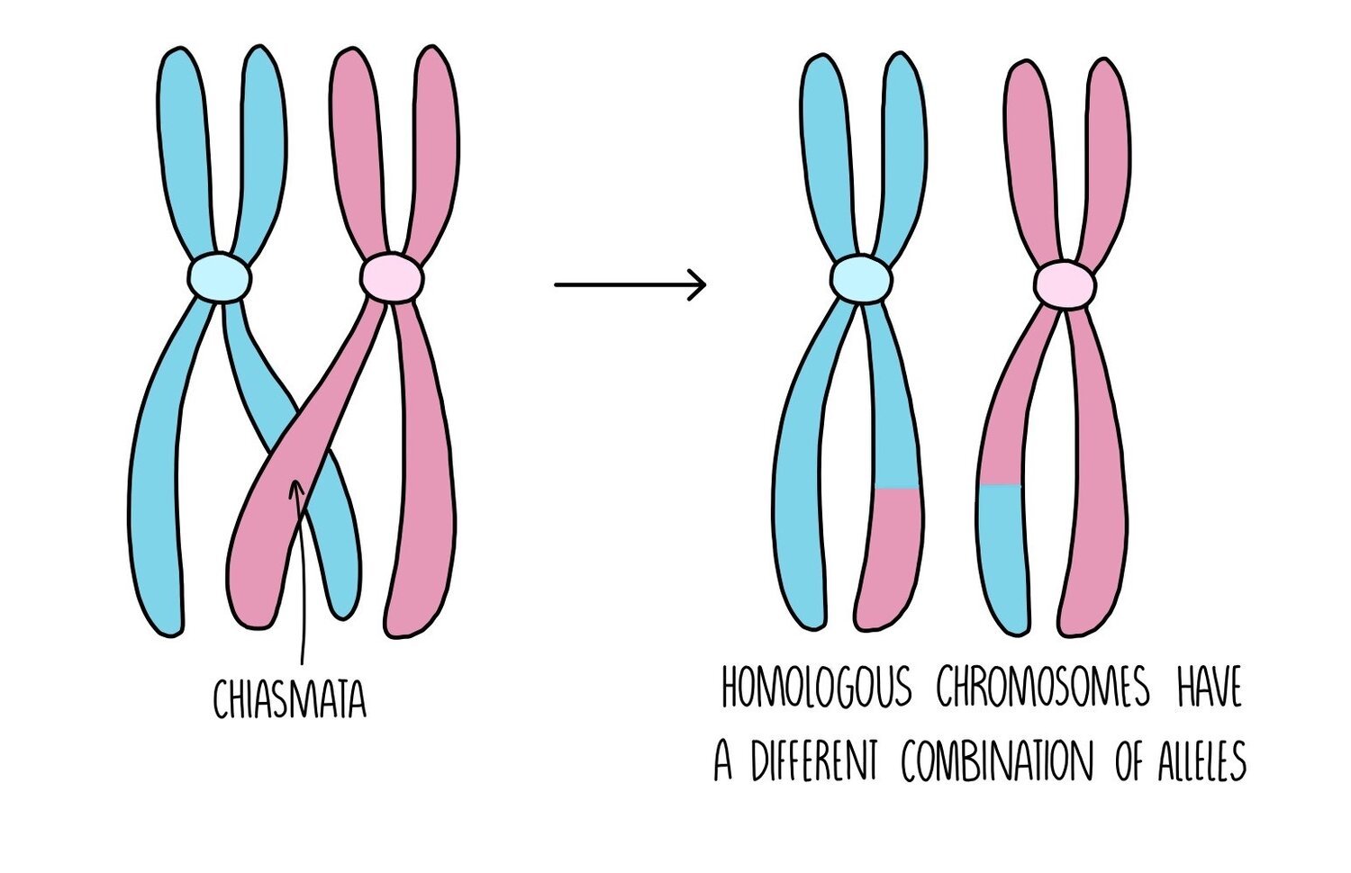
During prophase I of meiosis, a process called crossing over occurs. This is when the homologous chromosomes move towards each other and exchange genetic material. A chromatid from the maternal chromosome becomes twisted around the paternal chromosome and they connect through a structure called the chiasmata. Pieces of chromosomes are exchanged and the chromatids separate, forming chromosomes with different combinations of alleles.
Independent assortment
Depending on the order in which chromosomes line up along the equator of the cell during metaphase, different combinations of chromosomes will end up in each gamete. The way in which the chromosomes align themselves on the spindle fibre is completely random, resulting in a huge number of possibilities of chromosomal combinations in the gametes.
Stem Cells and Potency

Stem cells are unspecialised cells which have the ability to become specialised cells, such as heart cells or neurons. The process by which a stem cell is converted from an unspecialised cell to a specialised cell is called cell differentiation. Stem cells have an unlimited capacity to divide and can produce lots more stem cells by mitosis.
The ability of stem cells to undergo differentiation is referred to as potency - there are different levels of potency:
Totipotent - totipotent cells have the ability to divide into any type of cell (including the extraembryonic cells which make up the placenta and umbilical cord).
Pluripotent - pluripotent cells can divide into any type of cell except the extraembryonic cells.
Multipotent - these cells can divide into a handful of different cell types
Unipotent - these cells can only divide into one type of cell
Adult bone marrow contains multipotent adult stem cells which can divide and differentiate to replace old blood cells. They are responsible for forming all the different types of red and white blood cells. In plants, stem cells are present in regions called meristems, found at the tip of the shoot and roots. These have greater potency than adult stem cells, and can divide to form almost any kind of cell.
Stem cells in medicine
Stem cells are being used as a treatment for certain diseases:
Stem cell transplants are given to patients with leukaemia - leukaemia is a type of cancer which destroys stem cells so bone marrow transplants are used to replace the lost stem cells.
Research is being carried out to develop ways of growing whole organs from stem cells. The organs can then be transplanted into the patient to replace organs that have been damaged or are diseased e.g. pancreatic transplants can be given to people with diabetes. This approach will help those who currently have to wait years for an organ donation.
Levels of organisation

The cell is the ‘basic building block of life’ and is the smallest functioning part of an organism. A group of cells working together is called a tissue and a collection of tissues all performing a specific function is called an organ. Multiple organs which are connected together are referred to as an organ system.
Examples of organs include the heart and lungs (in animals) and leaves and roots (in plants).
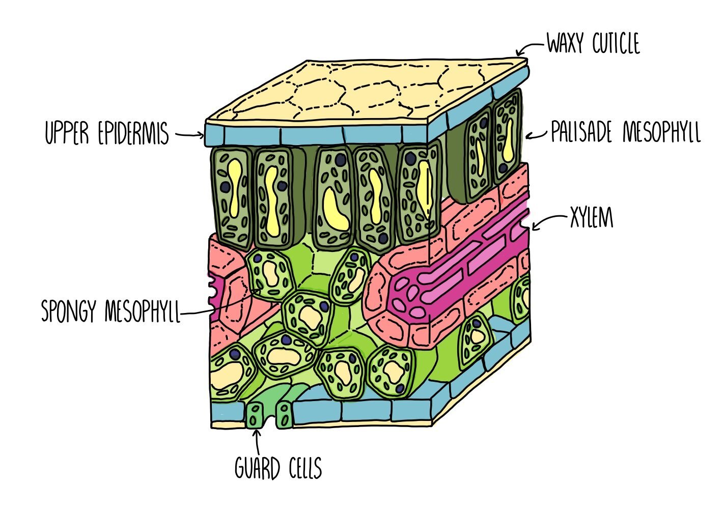
The leaf is an example of a plant organ and is made up of the following tissues:
Upper epidermis – covered in a waterproof waxy cuticle to reduce transpiration.
Palisade mesophyll – tightly packed cells filled with mitochondria, located towards the top of the leaf to absorb as much light as possible. This is where most photosynthesis takes place.
Spongy mesophyll – loosely arranged cells. Air spaces allow circulation of gases for gas exchange.
Phloem – transport of sugars and amino acids (translocation).
Xylem – transport of water and mineral ions (transpiration).
Lower epidermis – contains stomata which open and close to allow gas exchange to take place.
The lungs are an example of an organ found in animals and is made up of the following tissues:
Endothelium – forms the capillary walls which supply alveoli with oxygen and nutrients.
Fibrous connective tissue – helps to push air out of the lungs during exhalation.
Squamous epithelial tissue – makes up the walls of the alveoli.
Examples of organ systems include the respiratory system, circulatory system, reproductive system and digestive system.