Sexual Reproduction and Meiosis
Gametes
Gametes are sex cells (the sperm and egg in humans). Gametes are haploid which means they contain half the number of chromosomes as the rest of the cells which make up our body. This means that when two gametes fuse during sexual reproduction, the fertilised egg (called a zygote) contains the full number of chromosomes i.e. it is diploid. In humans, the diploid number of chromosomes is 46 (23 pairs), which means that gametes contain just 23 chromosomes.
During sexual reproduction, the nucleus of the sperm cell fuses with the nucleus of the egg cell - this fusion of nuclei is called fertilisation.
Adaptations of gametes
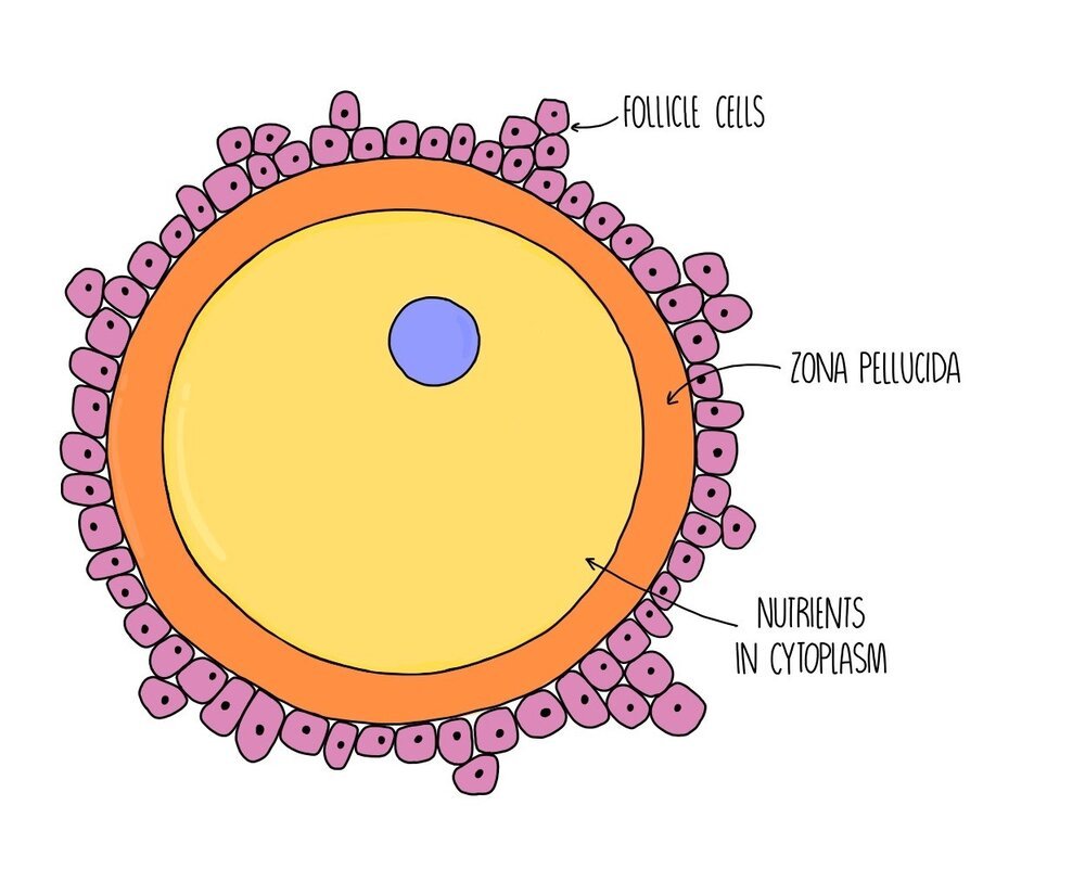
Egg cells are specialised for fertilisation in the following ways:
Cytoplasm contains nutrients for growth of the developing embryo.
There is an outer layer called the zona pellucida - this changes after fertilisation so that no further sperm can penetrate.
Follicle cells outside the zona pellucida form a protective coating around the egg cell.
Sperm cells are also specialised to carry out fertilisation:
Overall streamlined shape for faster swimming.
Tail (flagellum) enables the sperm to swim.
Contain lots of mitochondria to provide ATP for movement.
The head of the sperm contains the acrosome - this is filled with digestive enzymes which break down the egg’s zona pellucida and allow the sperm to reach the egg cell membrane.

The Acrosome Reaction
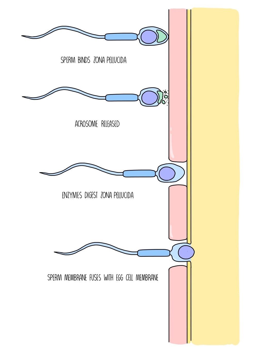
When a sperm reaches the egg cell, it binds to its outer layer (the zona pellucida) through attachment to sperm-binding proteins. Once bound, it releases the digestive enzymes that are contained in the acrosome. These enzymes digest a tunnel through the zona pellucida so that the sperm can reach the plasma membrane of the egg cell. The plasma membranes of the sperm cell and the egg cell fuse and the sperm releases its nucleus into the egg cell cytoplasm. The nuclei of the sperm and egg fuse - fertilisation has taken place and a zygote is formed.
The Cortical Reaction
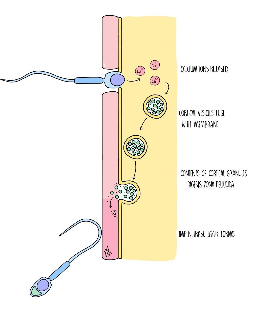
The fusion of the sperm cell and egg cell membranes triggers the release of calcium ions which stimulates vesicles containing cortical granules to move and fuse with the egg cell membrane. The cortical granules are released into the zona pellucida, which continue breaking down the zona pellucida, removing the remaining sperm-binding proteins so that no further sperm can bind. Other substances within the cortical granules produce a new outer layer which is thick and impenetrable to sperm cells. This process prevents multiple sperm cells from fertilising the egg, which would result in the zygote having an abnormal number of chromosomes.
Meiosis
Meiosis is the type of cell division which produces gametes for sexual reproduction. Unlike mitosis, the daughter cells are genetically different from the parent cell and contain just half the number of chromosomes (i.e. they are haploid). When two haploid gametes join during fertilisation, a diploid cell called a zygote is formed. Meiosis involves two rounds of cell division which are referred to as meiosis I and meiosis II.
It takes place in the following stages:
Interphase: the DNA replicates so there are now two identical copies of each chromosome (referred to as chromatids).
Meiosis I
Prophase I: chromatids condense and arrange themselves into homologous pairs (called bivalents). Crossing over occurs (see below). The nuclear envelope disintegrates and spindle fibres form.
Metaphase I: homologous chromosomes line up along the equator and attach to the spindle fibre by their centromeres.
Anaphase I: homologous chromosomes are separated
Telophase I: chromosomes reach opposite poles of the cell. Nuclear envelope reforms around the chromosomes. Cytokinesis results in the formation of two daughter cells.
Meiosis II
Prophase II: chromosomes condense, nuclear envelope disintegrates and spindle fibres form.
Metaphase II: chromosomes attach to the spindle fibre by their centromeres.
Anaphase II: sister chromatids are separated.
Telophase II: chromatids reach opposite poles of the cell. Nuclear envelope reforms and cytokinesis takes places. Four genetically unique daughter cells are produced.

Meiosis increases genetic variation
From an evolutionary point of view, it is important that organisms produce offspring that show as much genetic variation as possible. Imagine if a mother duck gave birth to a group of ducklings that were all had very similar genes - these ducklings will all be equally vulnerable to the same diseases and other threats to their survival. Meiosis increases genetic variation in two ways - crossing over and independent assortment.
Crossing Over
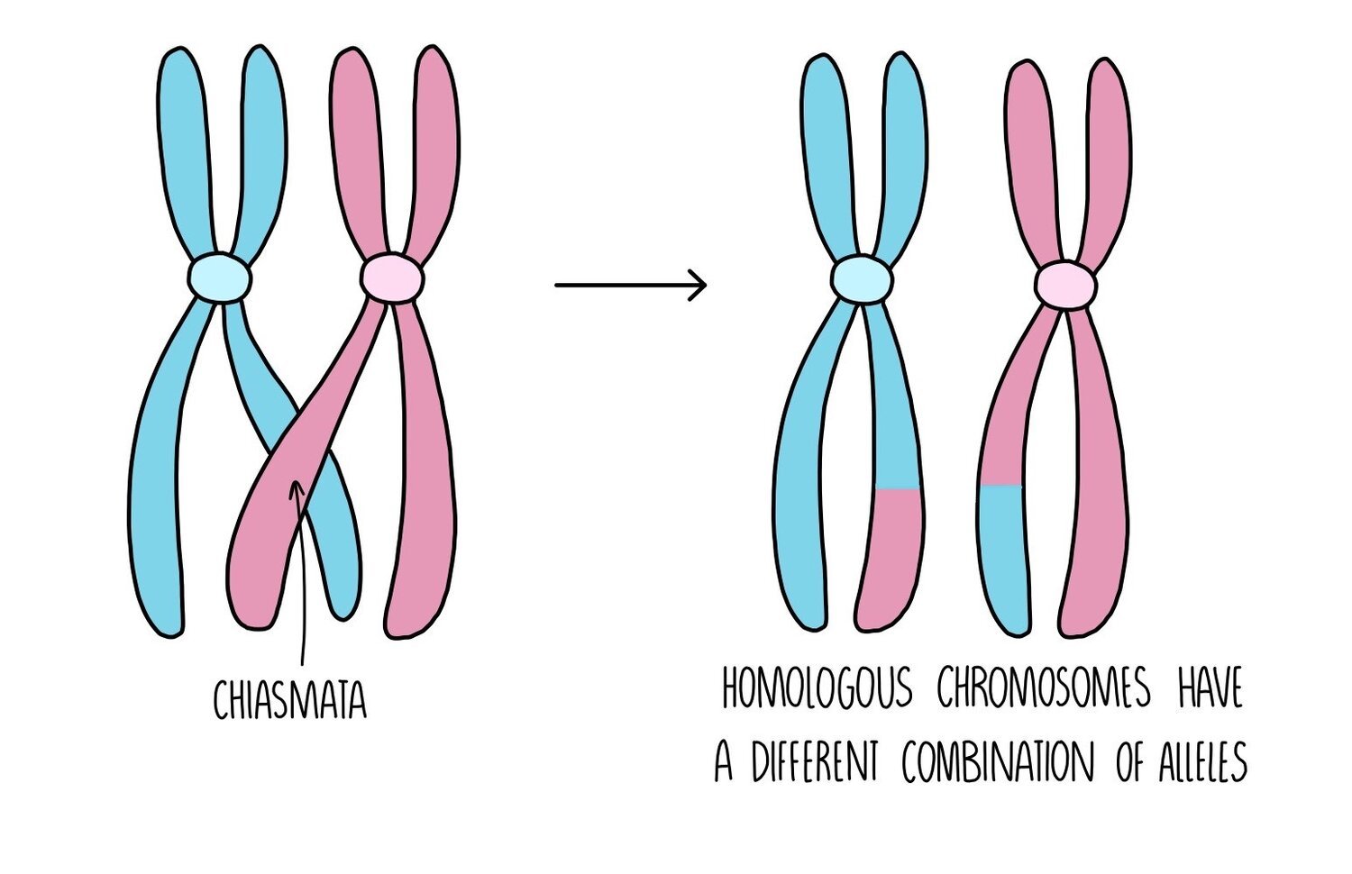
During prophase I of meiosis, a process called crossing over occurs. This is when the homologous chromosomes move towards each other and exchange genetic material. A chromatid from the maternal chromosome becomes twisted around the paternal chromosome and they connect through a structure called the chiasmata. Pieces of chromosomes are exchanged and the chromatids separate, forming chromosomes with different combinations of alleles.
Independent assortment
Depending on the order in which chromosomes line up along the equator of the cell during metaphase, different combinations of chromosomes will end up in each gamete. The way in which the chromosomes align themselves on the spindle fibre is completely random, resulting in a huge number of possibilities of chromosomal combinations in the gametes.
Linked genes

The position of a gene on a chromosome is called its locus. If the loci of two different genes are on the same chromosome, they are likely to be inherited together and are said to be linked. The only way that the genes will not be inherited together is if crossing over separates them during meiosis (the chiasmata would have to form between the two genes). The closer the loci of the two genes, the less likely this is to happen and the higher the probability that the genes will be inherited together. This means that any offspring will probably express both phenotypes together than either phenotype separately.
The image below shows an example of gene linkage in rats. If the genes for coat colour and eye colour are located on the same chromosome, they will be inherited together and the offspring will show both phenotypes together. For example, there will be rats with both black fur and red eyes, and with both brown fur and white eyes but not many with black fur + white eyes (or likewise, brown fur + red eyes). To get these combinations of phenotypes, crossing over must have occurred between the homologous chromosomes to cause the alleles to end up on the same chromosome and be inherited together.
Sex Linkage

Genes which are located on one of the sex chromosomes (X or Y) are said to be sex-linked and their expression will depend on whether the offspring is male (XY) or female (XX). The Y chromosome is much smaller than the X chromosome, so most alleles are carried on the X chromosome (they are X-linked). Men only have one X chromosome which means that they will only inherit one allele for these genes, compared to women who will inherit two. This means that if men inherit a recessive allele (which causes disease) for a gene found on the X-chromosome, they will have the disease. Women who inherit the recessive allele will just be a carrier, since they have another X chromosome with the dominant, functioning allele. For women to have X-linked diseases, they must inherit two disease alleles (they will have a homozygous recessive phenotype). Examples of sex linked disorders include haemophilia and red-green colour blindness.