The Nervous System
How the nervous system works
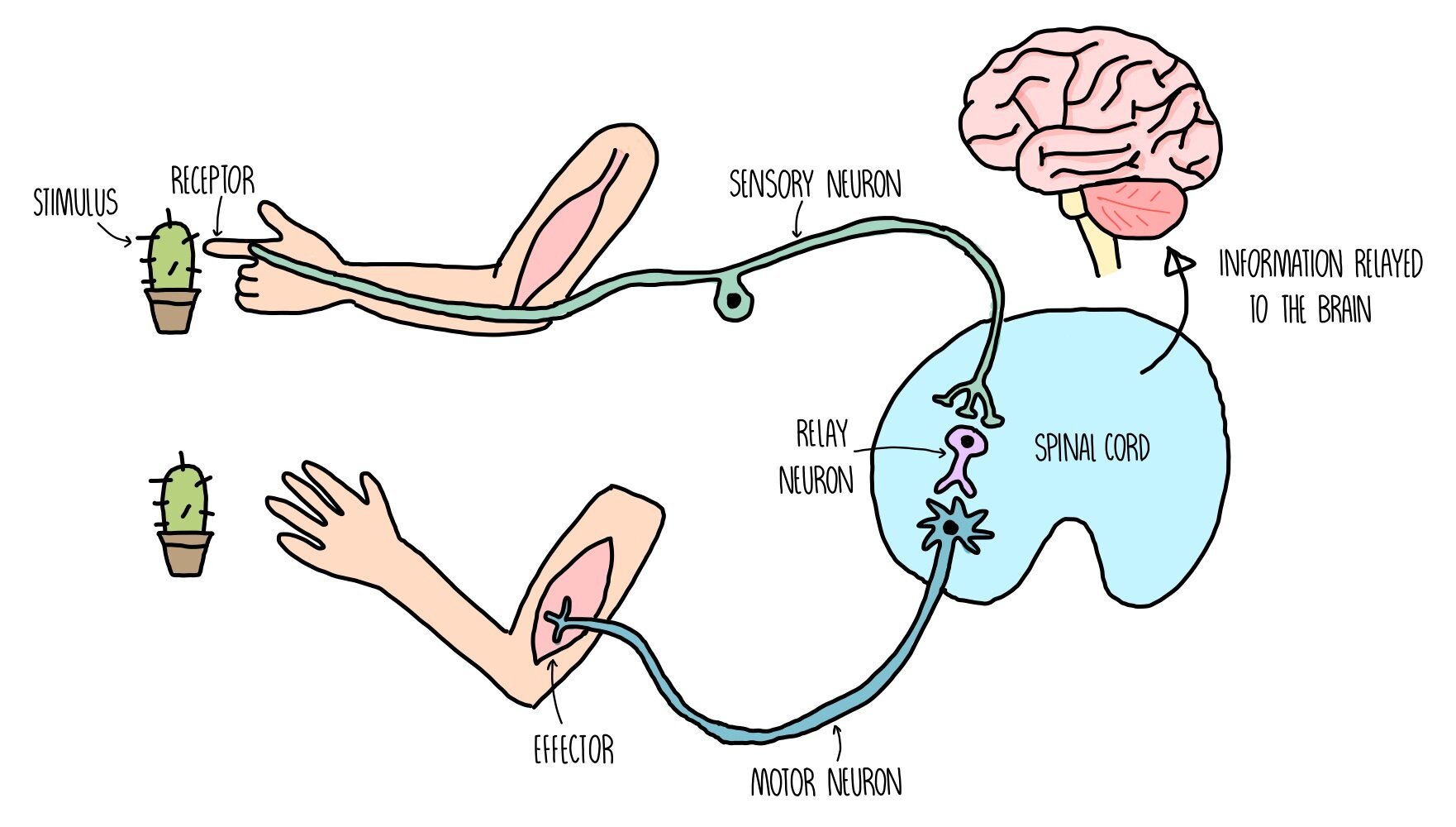
The nervous system detects changes in our environment (known as stimuli) through cells called receptors.
Receptors are sensitive to a number of different aspects of our environment, such as light, pressure (touch) and chemicals in the air (smell).
When receptors detect certain stimuli, they signal to the central nervous system (CNS) through initiating an electrical impulse through a neuron (nerve cell).
The neuron which sends an electrical impulse from the receptor within the sense organ and the coordination centre is called the sensory neuron.
The coordination centre receives impulses from various receptors around the body, processes the information and coordinates a response by signalling to other parts of the body. Coordination centres include the brain, spinal cord and pancreas.
These organs will signal to an effector (a muscle or gland) by releasing an electrical impulse along a motor neuron.
Stimulation of an effector will produce a response such as muscle contraction or hormonal release.
Receptors
Our nervous system uses receptors to detect stimuli (changes in the environment) and pass on this information to the CNS. Receptors can either be whole cells (e.g. photoreceptors are cells which are sensitive to light) or proteins molecules which are found on the cell surface membrane. Each receptor is specific to a single type of stimulus, such as light, temperature or glucose concentration. When a receptor is not stimulated, there is a charge difference between the inside and outside of the membrane and it is said to be polarised. When the receptor detects a stimulus, the permeability of its cell membrane changes which changes the charge difference (potential difference) across the membrane. If the change in potential difference is large enough (i.e. it exceeds the threshold level), it will trigger an action potential (an electrical impulse) in a sensory neuron.
We contain the following receptors in our sense organs:
Chemoreceptors - receptors which detect chemicals
Thermoreceptors - receptors which detect heat
Mechanoreceptors - receptors which detect pressure (see the Pacinian corpuscle below)
Photoreceptors - receptors which detect light (e.g. rods and cones)

Pacinian corpuscles
Pacinian corpuscles are receptors which respond to changes in pressure - they are a type of mechanoreceptor. They are found deep in the skin and are abundant in the feet, fingers, external genitalia and in our joints. The Pacinian corpuscle consists of a single sensory neurone, surrounded by layers of tissue which are each separated by a gel, forming an onion-like structure.
The Pacinian corpuscle contains stretch-mediated sodium ion channels in the cell surface membrane. Under normal conditions these channels are closed but when pressure is applied these channels become deformed and open, allowing a rapid influx of sodium ions. This makes the membrane potential in the neurone less negative (depolarisation), producing a generator potential which can then produce an action potential.
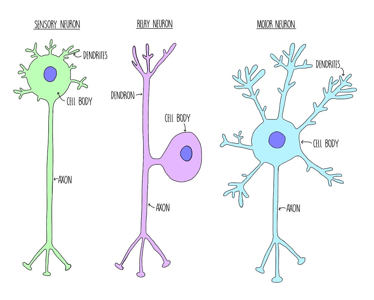
Types of neurone
Neurones are cells which carry information to and from the central nervous system, in the form of electrical impulses called action potentials. There are three different types of neurone, with slightly different structures. What they all have in common, however, is a cell body containing a nucleus, dendrites which carry an action potential towards the cell body and an axon which carries the action potential away from the cell body.
Sensory neurones carry action potentials from receptors to the central nervous system. They consist of one long dendron and a short axon.
Relay neurons carry action potentials between the sensory and motor neurons and are found within the CNS. They have lots of short dendrites.
Motor neurones carry action potentials from the CNS to an effector. They have lots of short dendrites and one long axon.
Resting potential
When a neurone is not firing (i.e. it is not transmitting an action potential), there is a difference in charge between the inside and the outside of the membrane (it is polarised). This charge difference is referred to as the resting potential and is usually around -70 mV. Polarisation of neuronal cell membranes at rest occurs due to the action of sodium-potassium ion pumps. These pumps are found within the cell membrane and actively transport sodium and potassium ions into and out of the neurone. For every three sodium ions that the proteins pump out of the cell, they pump two potassium ions into the cell. This ensures that there are always more positive ions out of the cell compared to inside the cell and makes sure there is a charge difference across the membrane.

Action potential
When a neurone is stimulated, the charge difference between the inside and outside of the cell membrane is lost and the membrane is depolarised. If enough charge is lost and depolarisation exceeds -55 mV, an action potential will occur in that neurone. The -55 mV ‘limit’ is known as the threshold potential - any depolarisation above this number will result in an action potential whereas anything less than that will result in nothing. We therefore refer to action potentials as an “all-or-nothing” response.
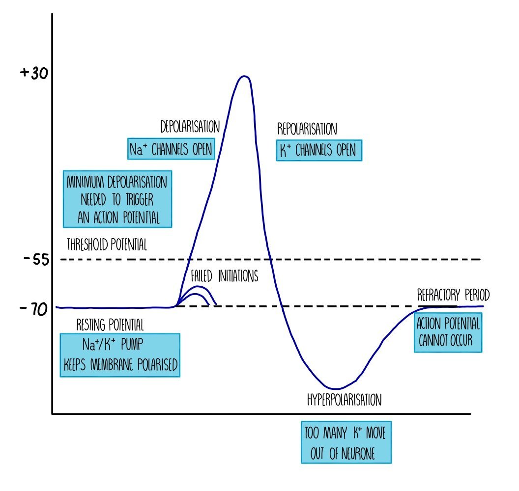
Depolarisation during an action potential occurs because sodium ion channels open up in the membrane. Remember that the sodium-potassium ion pump has been actively transporting sodium ions out of the neurone, creating a sodium ion concentration gradient. This means that when sodium ion channels open, sodium ions flood into the neuron by facilitated diffusion. The potential difference across the membrane is reduced until it reaches a voltage of around +30 mV.
Sodium ion channels close and potassium ion channels open, which causes potassium ions to move out of the neurone down their concentration gradient. The movement of positive ions out of the cell means that there is a charge difference again across the membrane - this is called repolarisation. However, the charge difference exceeds the resting potential and becomes ‘hyperpolarised’. This is because the potassium ion channels are slow to close and too many potassium ions diffuse out of the neurone. The action of the sodium-potassium ion pump restores the balance between sodium and potassium ions on either side of the membrane and returns the neurone to its resting potential of -70 mV.
Immediately after an action potential is a brief period called the refractory period. During the refractory period, the neurone cannot be stimulated and an action potential cannot occur. This is because the ion channels are recovering and they cannot be made to open. The refractory period is important because it ensures that action potentials do not overlap (i.e. they pass along the neurone as separate impulses) and that action potentials are unidirectional.
Once an action potential occurs in one part of the neuron, it will stimulate an action potential in the adjacent part of the neuron, creating a kind of ‘Mexican wave’ of depolarisation. This wave of depolarisation occurs because the sodium ions which diffuse into the neuron diffuse sideways, causing voltage-gated ion channels in the next portion of the neurone to open, so sodium ions move into the neurone further along the membrane. The wave moves away from the part of the neurone which has just fired an action potential because that part of the neurone will be in the refractory period and cannot be stimulated.
Saltatory conduction
Some neurones are insulated with a fatty layer along the axon. This fatty layer is called a myelin sheath and is made up of a type of cell called a Schwann cell. The myelin sheath acts as an electrical insulator, which means that ions cannot move into or out of the myelinated portions of the neurone. However, there are gaps in the myelin sheath called nodes of Ranvier, where sodium ion channels and potassium ion channels are concentrated. Action potentials occur only at the nodes of Ranvier - when one node is stimulated this triggers depolarisation of the next node, causing the impulse to ‘jump’ from node to node. This type of nervous transmission is called saltatory conduction and is much faster than transmission along non-myelinated neurones, where the action potential has to travel along the entire length of the neurone in a wave of depolarisation. The speed at which an action potential moves along a neurone is known as the conduction velocity - the higher the conduction velocity, the faster the action potential is travelling. This means that action potentials along myelinated neurones have a higher conduction velocity compared to those travelling along non-myelinated neurones.
Size of the stimulus
An action potential is an ‘all-or-nothing’ response. If the threshold potential is reached, an action potential will occur. This action potential is always of the same voltage (depolarisation to +30 mV) regardless of whether the stimulus that initiated the action potential is small (e.g. a pinprick) or large (e.g. a sledgehammer). If the threshold isn’t reached then an action potential will not be fired. The difference between action potentials resulting from stimuli of different sizes is the frequency that action potentials are firing - the bigger the stimulus, the more often an action potential will occur along the neurone.
Myelination
Some neurones are insulated with a fatty layer along the axon. This fatty layer is called a myelin sheath and is made up of a type of cell called a Schwann cell. The myelin sheath acts as an electrical insulator, which means that ions cannot move into or out of the myelinated portions of the neurone. However, there are gaps in the myelin sheath called nodes of Ranvier, where sodium ion channels and potassium ion channels are concentrated. Action potentials occur only at the nodes of Ranvier - when one node is stimulated this triggers depolarisation of the next node, causing the impulse to ‘jump’ from node to node. This type of nervous transmission is called saltatory conduction and is much faster than transmission along non-myelinated neurones, where the action potential has to travel along the entire length of the neurone in a wave of depolarisation. The speed at which an action potential moves along a neurone is known as the conduction velocity - the higher the conduction velocity, the faster the action potential is travelling. This means that action potentials along myelinated neurones have a higher conduction velocity compared to those travelling along non-myelinated neurones.
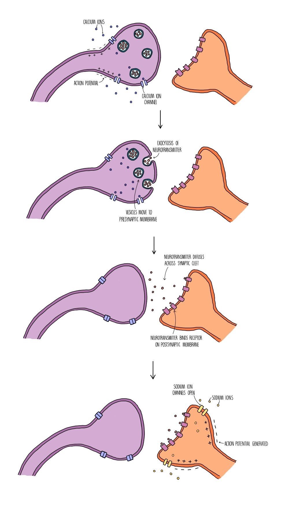
Synapses
A synapse is a gap found between neurones (or between a motor neurone and an effector). Electrical impulses cannot pass through the gap, so neurones release neurotransmitters from one neurone to the next to stimulate an action potential in the next neurone. The neurone before the synapse is called the presynaptic neurone and the one after the synapse is called the postsynaptic neurone. The space between them is called the synaptic cleft. This means that action potentials will travel along the presynaptic neurone, through the synaptic cleft (via neurotransmitters) then along the postsynaptic neurone. The presynaptic neurone has a swelling at the end which is called the synaptic knob.
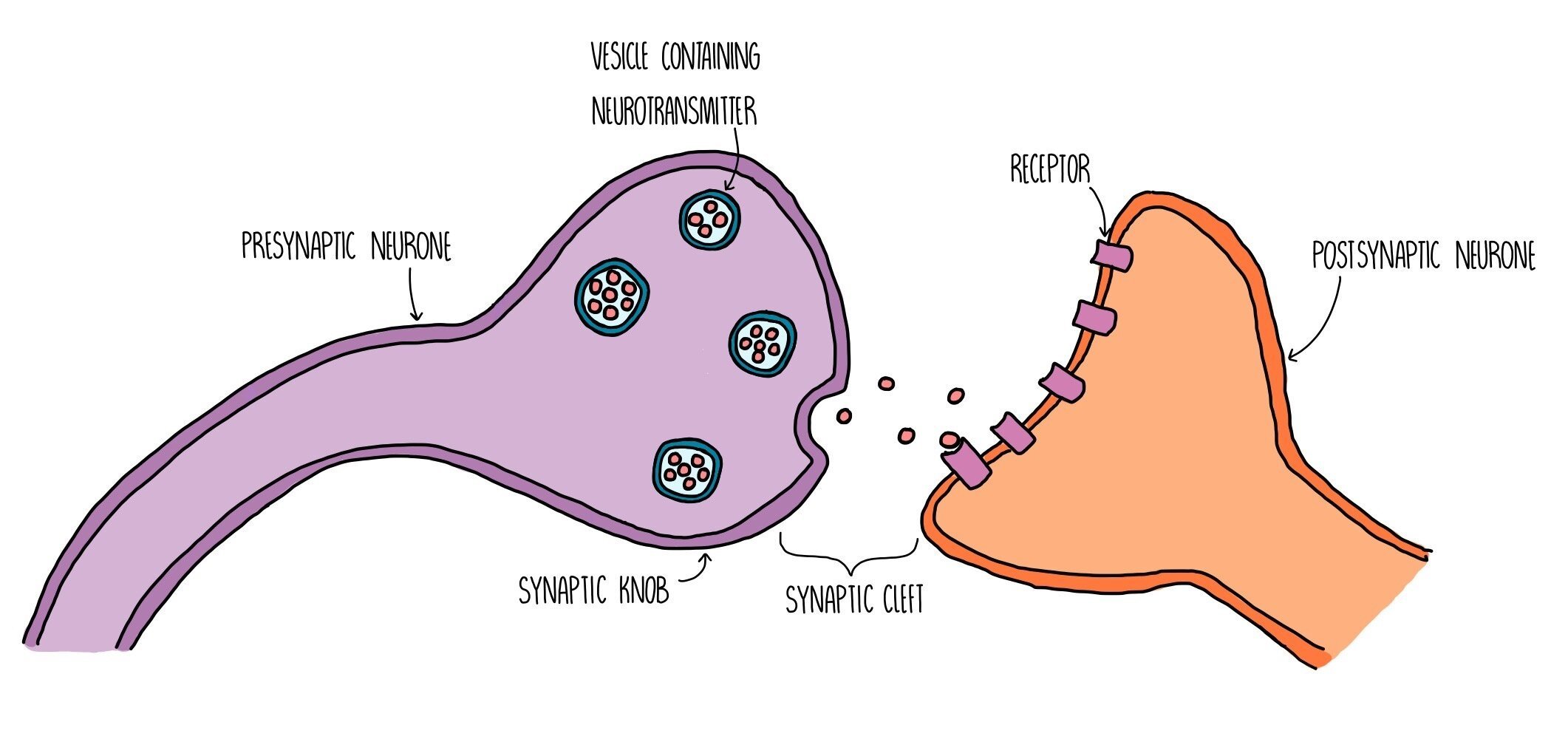
Synaptic transmission takes place in the following stages:
An action potential arrives at the end of the presynaptic neurone (at the synaptic knob) and triggers the opening of voltage-gated calcium ion channels.
Calcium ions move into the synaptic knob by facilitated diffusion and trigger the movement of vesicles containing neurotransmitters (such as acetylcholine or dopamine) towards the presynaptic membrane.
The vesicles fuse with the presynaptic membrane and their contents is released by exocytosis.
The neurotransmitters diffuse across the synaptic cleft and bind to specific receptors on the postsynaptic membrane.
This triggers the opening of sodium ion channels in the postsynaptic membrane. Sodium ions move into the postsynaptic neurone, causing depolarisation and triggering an action potential if the excitation exceeds the threshold potential of -55 mV.
The neurotransmitter is removed from the synaptic cleft which prevents the continuous stimulation of an action potential in the postsynaptic neurone. The neurotransmitter is either reabsorbed by the presynaptic neurone (and recycled) or broken down by enzymes in the synaptic cleft (and the products are reabsorbed).
Receptors which are complementary to neurotransmitters, such as acetylcholine, are only found on the postsynaptic membrane - there are none within the postsynaptic membrane). This ensures that the action potential always travels in one direction only (unidirectional) and prevents the nerve impulse from travelling backwards.
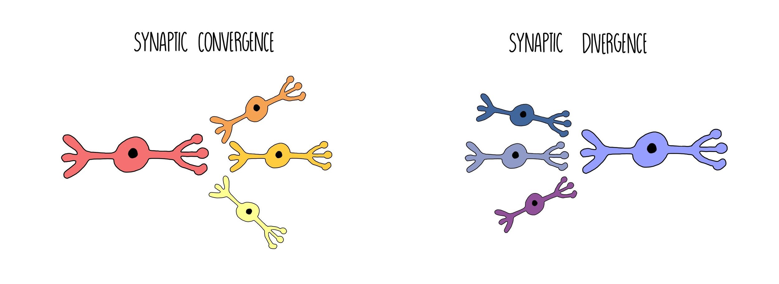
Synaptic divergence and convergence
When one neurone forms connections to multiple neurones through multiple synapses, the action potential can diverge, sending information to different parts of the body. This is known as synaptic divergence. On the other hand, multiple neurones can all connect to a single neurone, causing the action potentials from multiple neurones to converge and become amplified (synaptic convergence). This creates a stronger impulse when many different neurones are activated that form part of the same neural pathway.
Summation

Summation is when small amounts of neurotransmitter build up to trigger an action potential in the postsynaptic neuron. Spatial summation occurs when lots of presynaptic neurons converge on a single postsynaptic neuron. Although each of the presynaptic neurons are releasing small amounts of neurotransmitter (which alone would not be enough to exceed the threshold potential), the combined amount is enough to stimulate an impulse in the next neuron.
Temporal summation is when a single neuron fires action potentials in quick succession, repeatedly releasing neurotransmitter into the synaptic cleft. This causes the amount of neurotransmitter in the synaptic cleft to increase, making an action potential in the postsynaptic neuron more likely.
How the hormonal system works
The endocrine (hormonal) system is a group of glands, which secrete chemicals called hormones.
Glands secrete hormones when they are stimulated by neurons or by a change in concentration of a chemical substance (such as another hormone).
Hormones travel around our body in our bloodstream to their target organs where it produces an effect.
Target cells possess complementary receptors on their cell surface membranes that allow it to respond to the hormone, triggering a response inside the cell.
Hormones can be proteins/peptides e.g. insulin or steroids (a type of lipid) e.g. testosterone.
Compared to the nervous system, the endocrine system is:
Slower to bring about an effect
Longer-lasting
Widespread response (can act on multiple target tissues)
Involves the use of chemicals rather than electrical impulses

THE ENDOCRINE SYSTEM
Second messengers
Since hormones such as adrenaline, glucagon and insulin can’t get inside target cells to exert their effects directly, they have to use second messengers to activate reactions inside cells. For example:
Adrenaline binds to its receptor on the cell surface membrane of liver cells.
This activates the enzyme adenylyl cyclase, which catalyses the production of a second messenger called cyclic AMP (cAMP).
cAMP then activates a cascade of intracellular reactions.
For example, cAMP activates an enzyme called protein kinase A, which activates a cascade which eventually leads to the breakdown of glycogen (glycogenolysis).
The adrenal glands
The adrenal glands sit just on top of your kidneys and are made up of the medulla (in the middle) and the cortex (the outer part). The medulla and cortex secrete different hormones.
The medulla secretes hormones called catecholamines, which include adrenaline and noradrenaline. These hormones are released in times of stress and increase energy levels by:
Increasing heart rate
Increasing breathing rate
Converting glycogen into glucose (glycogenolysis)
Vasoconstriction of blood vessels to various organs, diverting blood flow to the brain and muscles.
The cortex secretes steroid hormones, including cortisol and aldosterone. These are also released in times of stress and have the following effects:
Suppression of the immune system
Increase blood pressure by stimulating the kidneys to take up more sodium ions and water.
Converting proteins and fats into glucose (gluconeogenesis), increasing the amount of glucose available for respiration.
The pancreas
The endocrine part of the pancreas is called the islets of Langerhans, clusters of cells that are surrounded by capillaries. The islets of Langerhans are made up of two types of cell: Alpha cells which secrete glucagon directly into the bloodstream and beta cells, which release insulin into the blood. Glucagon acts to increase blood glucose levels while insulin has the opposite effect.