The Respiratory System
Surface area to volume ratio
For aerobic respiration, organisms need to get oxygen and glucose to their cells and get rid of waste products (carbon dioxide and water). To maintain body temperature, organisms also need to transport heat.
Single-celled organisms like bacteria have a large surface area; a thin surface and a short diffusion pathway so do not need transport systems. They absorb gases by diffusion.
For large, multicellular organisms, their surface area: volume ratio is too small to get these substances by diffusion as there is less space for diffusion to take place.
Due to their size, it would take also too long for the substances to reach cells in the body’s centre (large diffusion pathway).
Therefore, multicellular organisms have mass transport systems (e.g. the circulatory system) and exchange organs (the lungs/intestine).
Adaptations of exchange surfaces
Most gas exchange surfaces are extremely thin (sometimes just one cell thick), ensuring a short diffusion pathway across the exchange surface. They will also have a large surface area to volume ratio which provides more space for the diffusion of gases. The organism itself will also have features which maximise the concentration gradient of gases across the exchange surface.
Adaptations of the lungs
Each alveolus is adapted to make gas exchange as efficient as possible:
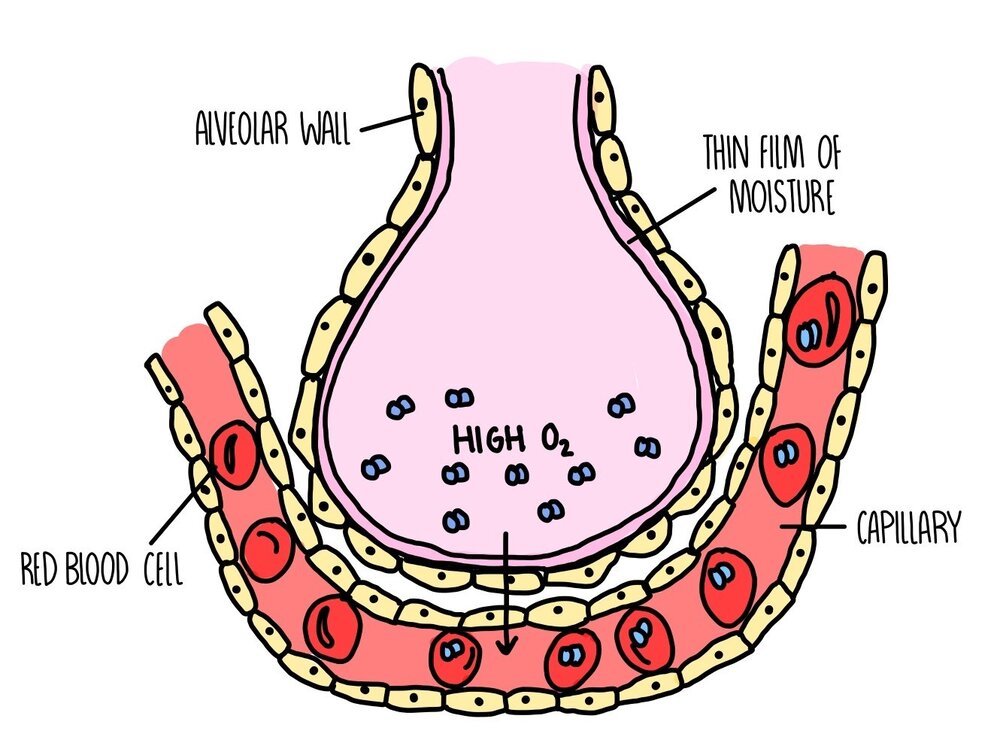
Large surface area: there are approximately 700 million alveoli in our lungs with a combined surface area of 70 square meters.
Good blood supply: lots of capillaries surround each alveolus
Short diffusion distance: the walls of both the alveoli and capillaries are just one cell thick
Moist surfaces: the liquid on the surface of alveoli dissolves gases and facilitates diffusion
Inhalation and exhalation: breathing in and out replaces the air in the alveoli, maintaining a steep concentration gradient.
Location in the centre of the body: the lungs are right in the middle of our body, inside our thorax. The core of our body is the warmest, giving the gas molecules more heat energy and making them move around faster (so quicker diffusion).
Respiratory system
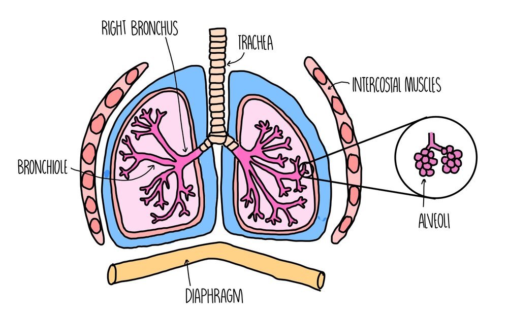
When we breathe, air enters our bodies through our nose and mouth and makes its way down the trachea. The trachea branches into two smaller tubes, called bronchi, which send air to each lung. The bronchi divide into even smaller tubes called bronchioles which finally send the air into air-sacs called alveoli. The alveoli look like a bunch of grapes and it’s this structure where gas exchange takes place. Oxygen diffuses from a region of high concentration in the alveoli to a region of low concentration in the bloodstream, where it travels to different tissues of the body and is used for respiration. Carbon dioxide travels in the other direction, from a region of high concentration in the bloodstream to a region of low concentration in the alveoli, where it travels up the trachea and is breathed out.
The lungs themselves are surrounded by the pleural membrane, a moist membrane which forms an airtight seal around the lungs. The ribcage protects the organs of the respiratory system and are surrounded by intercostal muscles and diaphragm, which move the rib cage during breathing to help bring air into or out of the lungs.
Structures in the human respiratory system have different functions:
Goblet cells – secrete mucus which traps dust and microorganisms to prevent infection. Found in the trachea, bronchi and larger bronchioles.
Cilia – beat to move mucus up towards the throat when it’s swallowed and destroyed by stomach acid. Found in the trachea, bronchi and bronchioles.
Elastic fibres – found in the walls of the trachea, bronchi, bronchioles and alveoli. The fibres stretch when we breathe in and recoil when we breathe out.
Smooth muscle – found in the trachea, bronchi and bronchioles. They control and relax to expand or narrow the airways.
Cartilage – found in rings in the walls of the trachea and bronchi. It’s strength and flexibility provides support and prevent the airways from collapsing when pressure drops during inhalation.
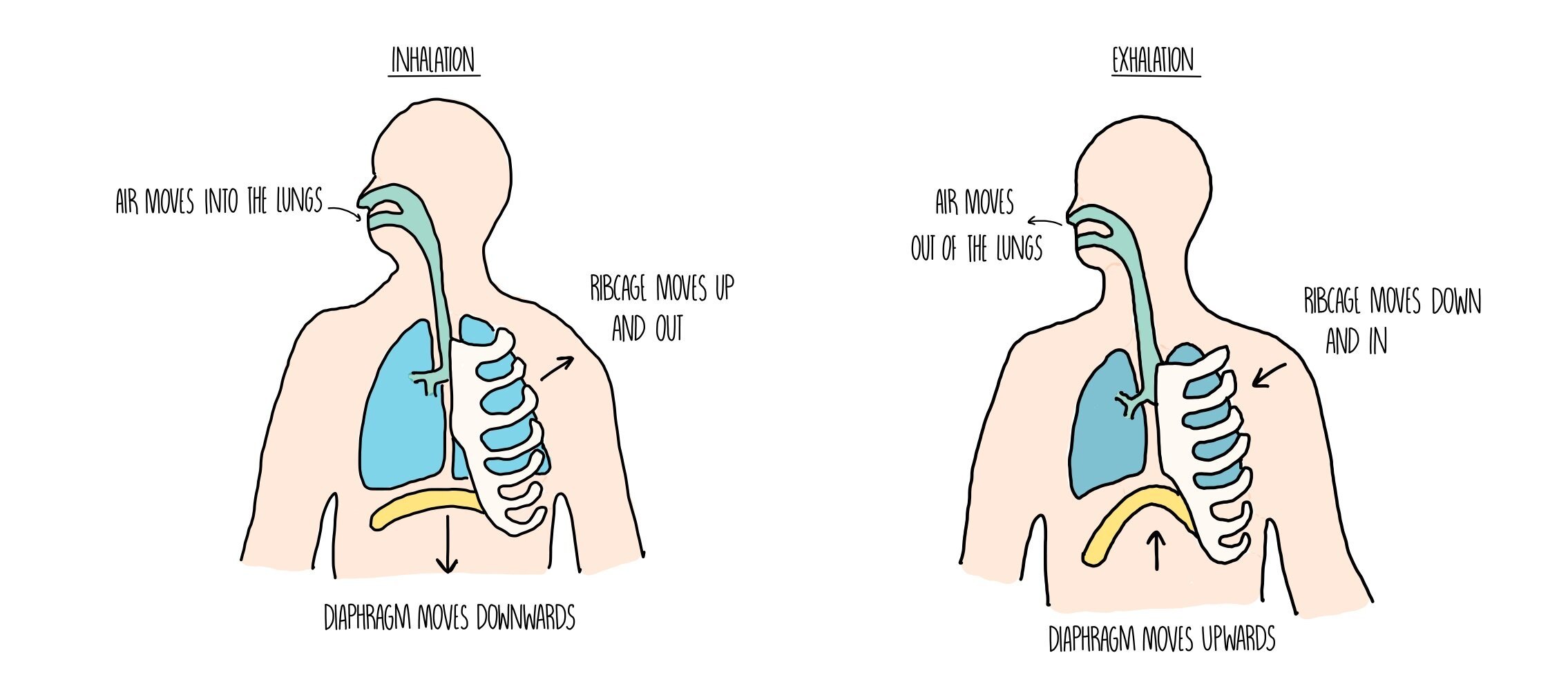
Inhalation and exhalation
During inhalation, the diaphragm contracts and flattens, while the rib cage moves up and out. This increases the volume of the thorax, resulting in a drop in pressure. The reduced pressure causes air to be sucked into our lungs.
During exhalation, the diaphragm relaxes and forms a dome shape, while the rib cage moves down and in. This reduces the volume of the thorax and increases the pressure, pushing air out of our lungs.
Investigating ventilation
You can investigate the volume of air that an individual is capable of breathing in and out using a piece of equipment called a spirometer. A spirometer consists of a chamber filled with oxygen and has a lid which moves up and down as a person breathes in and out. The spirometer is attached to a pen, which moves whenever the lid moves to draw a spirometer trace. A spirometer trace can be used to find the following information:
Tidal volume - this is the volume of air in each breath
Vital capacity - the maximum volume of air that a person can expel from the lungs after a maximum inhalation
Breathing rate - the number of breaths taken per minute
Oxygen consumption - the volume of oxygen used by the body
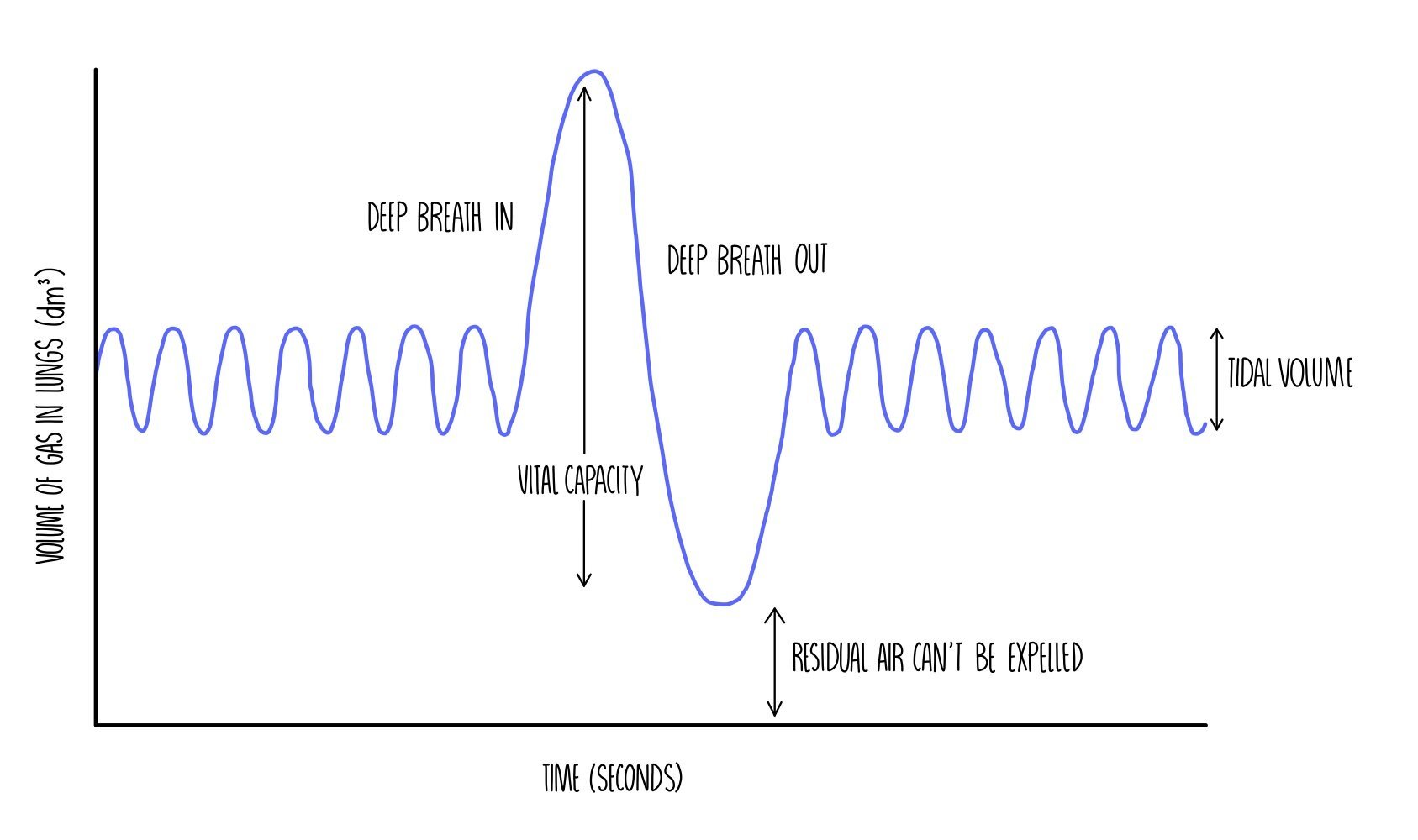
Spirometers contain a chemical called soda lime which absorbs any carbon dioxide that is breathed out. This means that the volume of gas in the chamber decreases as it is being used, since the oxygen in the chamber will be consumed by the person and the carbon dioxide that they exhale will be absorbed by the soda lime. Every time the spirometer is reused, the apparatus needs to be refilled with air.
Gas exchange in fish
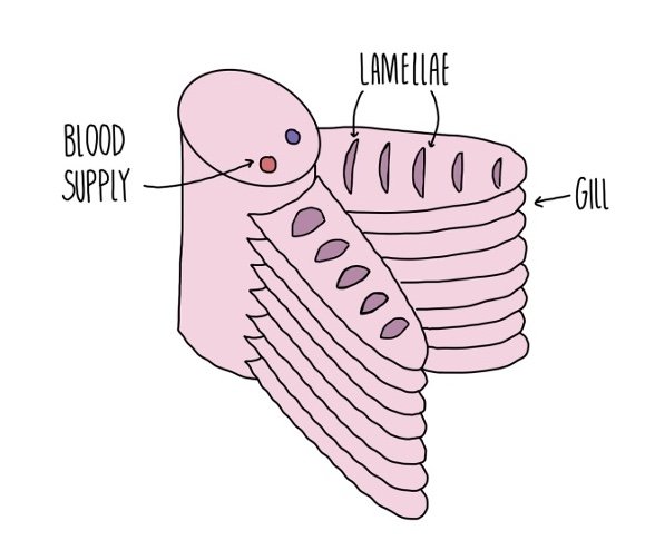
Water (containing oxygen) enters fish through the mouth and pass over the gills. Each gill is made up of several gill filaments stacked up on top of each other. The gill filaments are covered in lamellae to further increase their surface area. The lamellae have very thin walls to reduce the diffusion distance and are filled with capillaries.
Blood in the capillaries flows in the opposite direction to the water to maintain the concentration gradient of oxygen in the blood – this is known as the counter-current system.

Fish gills are ventilated in the following process:
When the fish opens its mouth, the floor of the bottom of the mouth (the buccal cavity) lowers, increasing the mouth’s volume.
Pressure drops inside the mouth, causing water to move through the mouth and into the buccal cavity.
When the fish closes its mouth, the floor of the buccal cavity moves up, reducing its volume.
Pressure increases, forcing water out of the cavity and across the gill filaments.
Each gill is covered by a protective bony flap called the operculum. When water moves over the gill filaments, it increases the pressure and forces the operculum on each side of the head to open. This allows water to leave the gills.
Gas exchange in insects
Insects have a tracheal system for gas exchange. Air enters the insect through pores in their outer surface called spiracles. Air then moves down the trachea, which branches off into a large number of tracheoles. The walls of the tracheoles are thin and porous, speeding up diffusion of gases to cells. Rhythmic abdominal movements push air into and out of the spiracles and maintain a steep concentration gradient.